Home
/ Anatomy Of Chest X Ray : Lateral Chest X Ray Anatomy Quotes | Chainimage : Living anatomy of the chest for 1st year medical students original version compiled by dr.
Anatomy Of Chest X Ray : Lateral Chest X Ray Anatomy Quotes | Chainimage : Living anatomy of the chest for 1st year medical students original version compiled by dr.
Anatomy Of Chest X Ray : Lateral Chest X Ray Anatomy Quotes | Chainimage : Living anatomy of the chest for 1st year medical students original version compiled by dr.. Common symptoms that can be diagnosed using chest. You have completed this module. In fact every radiologst should be an expert in chest film reading. Therefore, knowing the basics and pathologies in the ed setting is very important. Xray is a type of radiography and most widely used investigation.
It is almost always the first imaging study ordered to evaluate for pathologies of the thorax, although further diagnostic imaging, laboratory tests. In this article we will focus on: Many clinical conditions can be evaluated by this simple radiology test. In fact every radiologst should be an expert in chest film reading. Common symptoms that can be diagnosed using chest.
Chest X-ray anatomy from image.slidesharecdn.com Chest radiographs are the most common film taken in medicine. Note the larger appearing heart on the ap view. Heart and great vessels — assessment of the cardiovascular anatomy includes assessment of heart and chamber size as well as the position and size of the great. Many clinical conditions can be evaluated by this simple radiology test. Presence of metallic objects within the area of examination. Evaluation of a chest radiograph may appear to be simple, but is in fact a complex task requiring careful observation, sound understanding of chest anatomy, and knowledge of the principles of physiology and pathology. Common symptoms that can be diagnosed using chest. There are also important structures that are obscured or become visible.
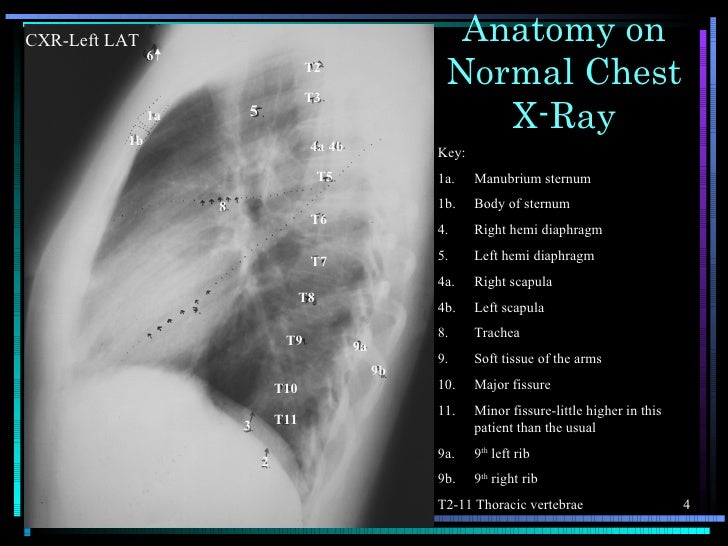
Labeled chest radiographs teaching radiologic anatomy with a level of detail appropriate for medical students.
Major structures are shown in fig. Both lungs should be well expanded and similar in volume. Each of these anatomical structures should be viewed using a systematic approach. Gillian lieberman forthe harvard 62. Note the larger appearing heart on the ap view. The interpretation of a chest film requires the understanding of basic principles. The interpretation of a chest film requires the understanding of basic principles. Labeled chest radiographs teaching radiologic anatomy with a level of detail appropriate for medical students. Therefore, knowing the basics and pathologies in the ed setting is very important. Consolidation, interstitial, nodule/mass, and atelectasis. A collection of anatomy notes covering the key anatomy concepts that medical students need to learn. Look for lung and pleural pathology. In fact every radiologst should be an expert in chest film reading.
It is almost always the first imaging study ordered to evaluate for pathologies of the thorax, although further diagnostic imaging, laboratory tests. Both lungs should be well expanded and similar in volume. The interpretation of a chest film requires the understanding of basic principles. Clinicalchest xray anatomy labeled clinical (i.redd.it). Chest radiographs are the most common film taken in medicine.
👨🏽💻Here's a recap of three posts on Chest X-Ray anatomy ... from i.pinimg.com Chest radiographs are the most common film taken in medicine. Consolidation, interstitial, nodule/mass, and atelectasis. Look for lung and pleural pathology. Gillian lieberman forthe harvard 62. Major structures are shown in fig. Evaluation of a chest radiograph may appear to be simple, but is in fact a complex task requiring careful observation, sound understanding of chest anatomy, and knowledge of the principles of physiology and pathology. Elbow anatomy anatomy bones upper limb anatomy radiology schools radiology student radiologic technology medical anatomy human anatomy and physiology medical coding. Common symptoms that can be diagnosed using chest.
Conclusion of living anatomy of the chest congratulations!
Living anatomy of the chest for 1st year medical students original version compiled by dr. The interpretation of a chest film requires the understanding of basic principles. Therefore, knowing the basics and pathologies in the ed setting is very important. Note the larger appearing heart on the ap view. Labeled chest radiographs teaching radiologic anatomy with a level of detail appropriate for medical students. Elbow anatomy anatomy bones upper limb anatomy radiology schools radiology student radiologic technology medical anatomy human anatomy and physiology medical coding. Evaluation of a chest radiograph may appear to be simple, but is in fact a complex task requiring careful observation, sound understanding of chest anatomy, and knowledge of the principles of physiology and pathology. Is there any inhaled foreign body? A collection of anatomy notes covering the key anatomy concepts that medical students need to learn. Consolidation, interstitial, nodule/mass, and atelectasis. Presence of metallic objects within the area of examination. It is almost always the first imaging study ordered to evaluate for pathologies of the thorax, although further diagnostic imaging, laboratory tests. Next, a good inspiration film should show at least the 10th or 11th posterior ribs.
Radiology basics of chest ct anatomy with annotated coronal images and scrollable axial images to help medical students and junior doctors learning anatomy. Labeled chest radiographs teaching radiologic anatomy with a level of detail appropriate for medical students. Next, a good inspiration film should show at least the 10th or 11th posterior ribs. Look for lung and pleural pathology. Chest radiographs are the most common film taken in medicine.
Labeled chest x-ray (CXR) | Cardiac anatomy, Radiology ... from i.pinimg.com You have completed this module. Therefore, knowing the basics and pathologies in the ed setting is very important. Conclusion of living anatomy of the chest congratulations! Labeled chest radiographs teaching radiologic anatomy with a level of detail appropriate for medical students. In this article we will focus on: There are also important structures that are obscured or become visible. Chest radiographs are the most common film taken in medicine. The interpretation of a chest film requires the understanding of basic principles.
Gillian lieberman forthe harvard 62.
Each of these anatomical structures should be viewed using a systematic approach. In fact every radiologst should be an expert in chest film reading. This imaging method can also check how a patient is responding to specific treatments. Legit, i can make out the trachea, aorta, outline of the heart, and the diaphragm. The interpretation of a chest film requires the understanding of basic principles. Xray is a type of radiography and most widely used investigation. Note the larger appearing heart on the ap view. Conclusion of living anatomy of the chest congratulations! Is there any inhaled foreign body? Abcde aproach comparison of pa vs. It is almost always the first imaging study ordered to evaluate for pathologies of the thorax, although further diagnostic imaging, laboratory tests. Gillian lieberman forthe harvard 62. You have completed this module.
It first appears too complicated to read the chest xrays because we barely know what anatomy of chest. Living anatomy of the chest for 1st year medical students original version compiled by dr.